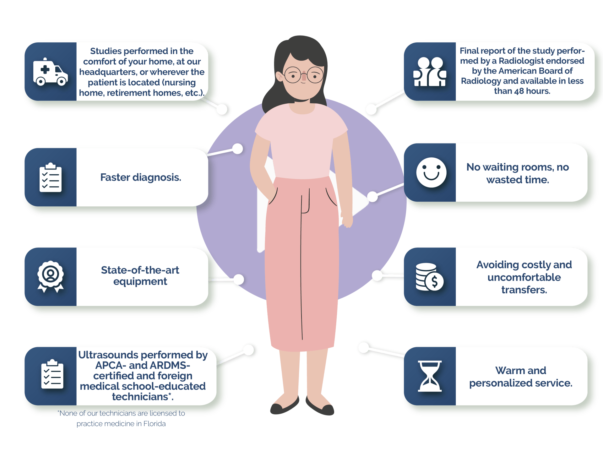
PATIENT SERVICES
We are the first mobile ultrasound service whose studies are performed by ultrasound technicians* certified by APCA (Alliance for Physician Certification & Advancement) and ARDMS ® (American Registry of Medical Sonography) who also have medical degrees* in other countries.
APCA and ARDMS ® are American institutions that accredit the academic training of doctors from other countries. Our strength is that we have an excellent group of professionals with extensive experience and training in performing all types of ultrasound, including echocardiography, arterial and venous
doppler, obstetric, gynecological, renal, abdominal, breast, thyroid, scrotal, soft tissue, among others. We are the difference in obtaining accurate, high quality and reliable studies, supported by the solid and specialized knowledge of our technical staff and our staff of certified radiologists, who are responsible for reporting the studies in a timely manner.
Complete Abdominal
Indicated for the study of internal organs in the abdomen, such as the liver, gallbladder, biliary tract, pancreas, spleen and kidneys, in order to determine possible causes of abdominal pain and/or distension, and also to correlate alterations of enzymes and/or markers in blood with pathological findings in the liver, pancreas and other organs. It also allows the diagnosis of infections and obstructions in the urinary tract, diagnosis and/or follow-up of known nodules or lesions, detection of congenital malformations, gallstones and kidney stones. It is also possible to assess some pathologies of the abdominal Aorta artery.
Preparation:
It is necessary to have fasting of six to eight hours before the study is performed.
Kidneys
It is a study in which the kidneys and urinary tract are evaluated to detect malformations, renal atrophy or hypertrophy, ectopic kidney, the presence and characterization of focal or diffuse lesions, detection of stones in both kidneys and urinary tract, and follow-up of known pathology. It may be necessary to complement the study with bladder ultrasound, as considered by the requesting physician.
Preparation: None.
THYROID
It is a neck study focused on the thyroid gland to detect nodules (palpable or not), determine their size, location and nature (solid, cystic or mixed). It is also indicated to correlate altered blood values of thyroid hormones and parathyroid hormone with pathological findings in the appearance of the thyroid and parathyroid gland.
Preparation: None.
Pelvic
It is indicated to study the female pelvic organs, such as the uterus, ovaries and tubes via transabdominal route.
It can be performed at any time, although sometimes it may be indicated on a certain day of the menstrual cycle. It is also useful in the detection of inflammatory and tumor lesions in the uterus, ovaries, tubes and other para adnexal structures. Among these lesions, the most common are ovarian cysts, uterine fibroids and endometrial hyperplasia. The early and often incidental finding of an ectopic pregnancy can in most cases save the life of the patient and hence the importance of its timely realization.
Preparation:
A full bladder is required.
OBSTETRIC
This test is performed transvaginally for the first 11 weeks, and preferably transabdominally after this time, unless it is necessary to evaluate the neck and placenta previa for which the transvaginal or translabial route is indicated. Useful to establish the presence of a live embryo/fetus, evaluate its situation, presentation and the development of its organs, estimate the gestation time of the pregnancy, diagnose congenital anomalies, as well as the evaluation of the placenta, cord and amniotic fluid depending on whether it is performed in the 1st, 2nd or 3rd trimester of gestation, in order to determine fetal growth and well-being.
Preparation: No preparation is required before 11 weeks, from 12 to 16 weeks it is required to have a full bladder, after 16 weeks it is not necessary to drink water prior to the study.
Gender reveal
Obstetric ultrasound performed to determine the sex of the baby at any time of pregnancy, it is usually done transvaginally between the 11th and 14th week, and transabdominal after that time.
If you want to know the sex of your baby, we are the ideal accomplices to make that moment magical.
Preparation: None.
Transvaginal
It allows the female pelvis to be explored transvaginally by placing a transducer in the vaginal canal. One of the benefits over the transabdominal route is that there is a better approach and a more accurate and detailed view of the gynecological organs. It can be performed at any time, although sometimes it may be indicated on a certain day of the menstrual cycle. It is also useful in the detection of inflammatory and tumor lesions in the uterus, ovaries, tubes and other para adnexal structures. Among these lesions, the most common are ovarian cysts, uterine fibroids and endometrial hyperplasia. The early and often incidental finding of an ectopic pregnancy can in most cases save the life of the patient, hence the importance of its timely performance. Another important contribution of this ultrasound is that it allows the evaluation of ovarian function through follicle count to complement the study of patients who are going to undergo fertilization therapies.
Preparation: None.
Liver and gallbladder
It is performed to detect any type of pathology in the liver, gallbladder and biliary tract. It allows to determine possible causes of a pain in the right upper quadrant of the abdomen, and also to correlate alterations in blood levels of liver tests with pathological findings found in liver, gallbladder and biliary tract, such as tumors, obstructive syndromes, inflammatory and/or infectious pathology, etc.
Preparation:
It is necessary to have fasting of six to eight hours before the study is performed.
Breast
It allows the study of the breast to detect and characterize breast nodules, as a complement to mammography, particularly in patients with dense breasts. It is also indicated in the evaluation of mastitis, palpable lumps, breast pain, in cases of secretions through the nipple, and as a preoperative study in aesthetic and reconstructive surgeries.
It is advisable to have previous mammograms and ultrasounds available on the day of the study.
Preparation: None.
Prostate
It is the study indicated to evaluate the prostate in men, its size and appearance to rule out or diagnose benign prostatic hyperplasia, detect possible nodules (with characteristics of malignancy and benignity), signs of inflammation and infection, alterations in the seminal vesicles.
Preparation: A full bladder is required.
Bladder
Study dedicated to evaluate the urinary bladder, including the thickness of the bladder wall, pre-micturition and post-micturition urinary volume, to check for adequate emptying. It allows the detection of tumors, polyps, diverticula, clots, sediment and bladder stones.
Preparation: A full bladder is required. In patients with bladder catheter, it is indicated to clamp the catheter with a non-cutting element to achieve urine retention.
Scrotal
It allows to observe and diagnose abnormalities in the testicles, scrotal sac and sperm ducts. It is used to verify the characteristics of a lump in the scrotum or testicles, determine injuries secondary to scrotal trauma, identify the causes of pain or increased volume of the testicles, determine the causes of sterility, locate a testicle that has not descended, monitoring of microcalcifications, among others.
Preparation: None.
Soft tissue
It is focused on the study of soft tissues such as skin, subcutaneous fatty tissue and musculoskeletal system. It allows detecting hernias, tumors, edema, joint effusions, collections, lymph nodes, location of foreign bodies (thorns, glass, metal, siliconomas, etc.), muscle tears, being able to clearly determine the size, location and nature (solid, cystic or mixed) of the findings, their extension and involvement of neighboring structures.
Preparation: None.
Neck and head soft tissue
Includes complete neck examination, including evaluation of the thyroid gland, salivary glands, ganglion chains and scalp for palpable lumps, thyroid nodules, thyroid hormone and parathyroid hormone disorders, alterations in salivation and other disorders.
Preparation: None.
Carotid
Also known as ultrasound of the neck vessels, in which the carotid and vertebral arteries on both sides are studied, it evaluates the blood flow of these vessels, allowing information to be obtained regarding vascular permeability, the direction of flow, presence and characterization of plaques, stenosis and vascular resistance. In addition, knowing the thickness of the carotid artery wall is a predictor of coronary artery disease in high-risk patients.
Preparation: None.
MSK
It allows the assessment of the structures of the musculoskeletal apparatus such as muscles, tendons, ligaments, bursae, and bone surfaces. The advantage of ultrasound over other diagnostic methods is that it is performed in real time and dynamically, so it is possible to observe, for example, the contraction of a muscle and the sliding of a tendon.
Preparation: No preparation is necessary for the correct performance of this ultrasound.
Preparation: None.
Baby Hip Joint
Study requested to evaluate the baby's hip before 3 months of age to determine whether or not there is congenital hip dysplasia in cases where it is suspected, and that according to the degree of severity can be classified as: dysplasia, subluxation and dislocation.
Preparation: None.
Echosonomovil benefits the patient
City skyline
The following ultrasounds are performed in ECHOSONOMOVIL
| STUDIO | SIRVE PARA EXAMINAR |
|---|---|
| Abdominal Ultrasound | Liver, spleen, gall bladder, pancreas, kidneys. |
| Bladder ultrasound | Urinary bladder |
| Prostate Ultrasound | Prostate |
| Gynecologic Ultrasound (pelvic or Transvaginal) | Uterus and ovaries |
| Breast Ultrasound | Mammary glands and armpits |
| Testicular Ultrasound | Testicles |
| Renal Ultrasound | Kidneys |
| Thyroid Ultrasound | Thyroid and parathyroid glands |
| Head and Neck Ultrasound | Neck nodes, salivary glands |
| Ultrasound - Soft Tissue Ultrasound | Palpable tumors in skin and subcutaneous cellular tissue |
| Ultrasound - Musculoskeletal | Musculoskeletal system |
| Ultrasound - Obstetrical | Pregnancy and fetus |
| Ultrasound baby hips | Hip joint |
| Echocardiogram (ECHO) | Heart and great vessels |
| Vascular doppler | Arteries, veins, testicles, tumors and vascular malformations |
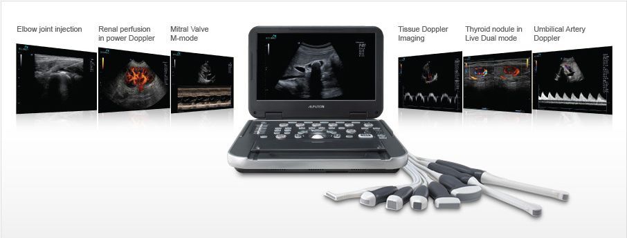
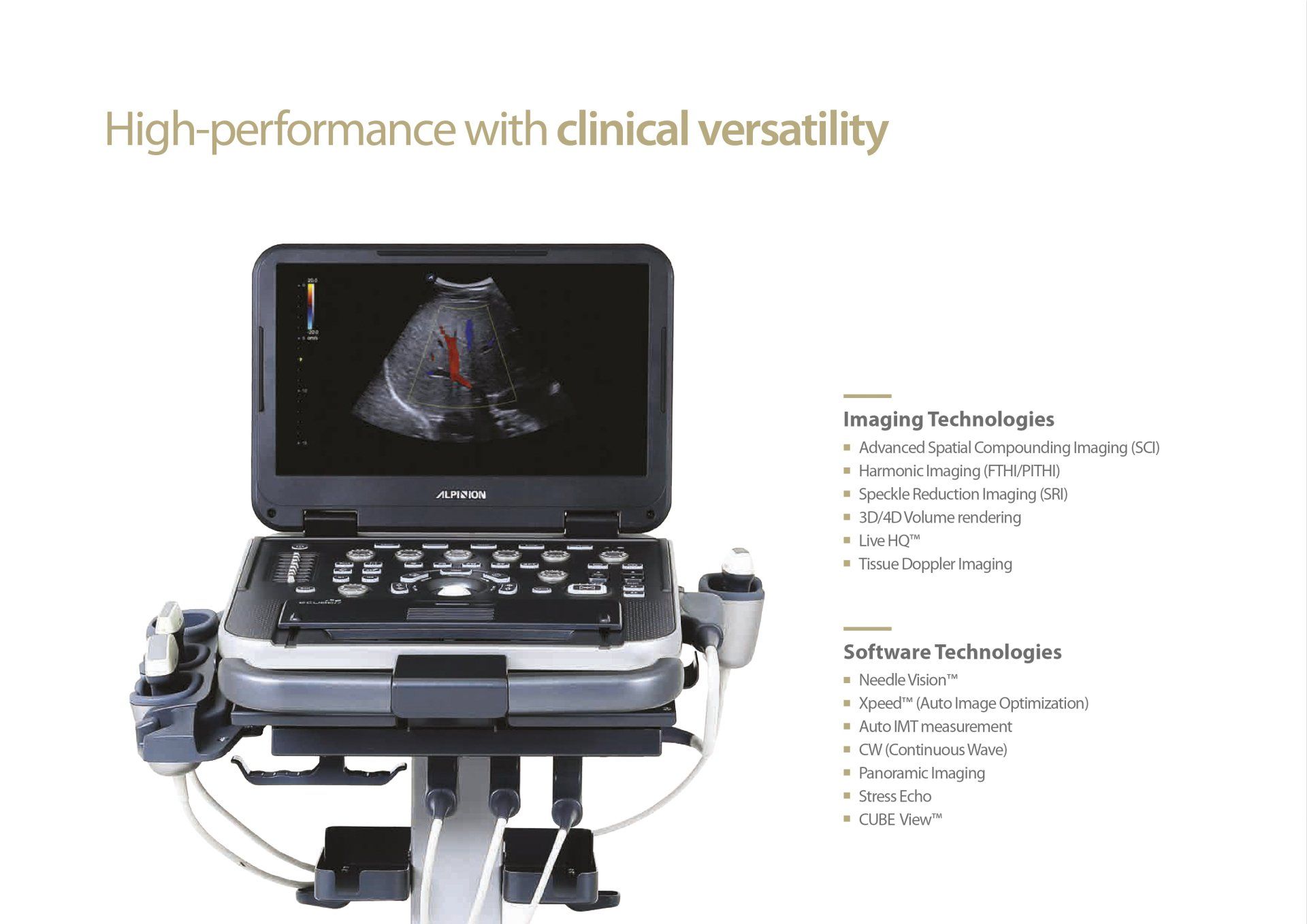
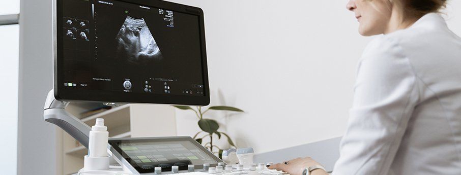
EQUIPMENT:
Modern ultrasound equipment is basically made up of 3 parts:
A) Transducer: device similar to a small microphone, which sends and collects the echo waves.
B) Monitor: It works just like a computer screen.
C) Computer: creates the images collected by the transducer to be viewed on the monitor.
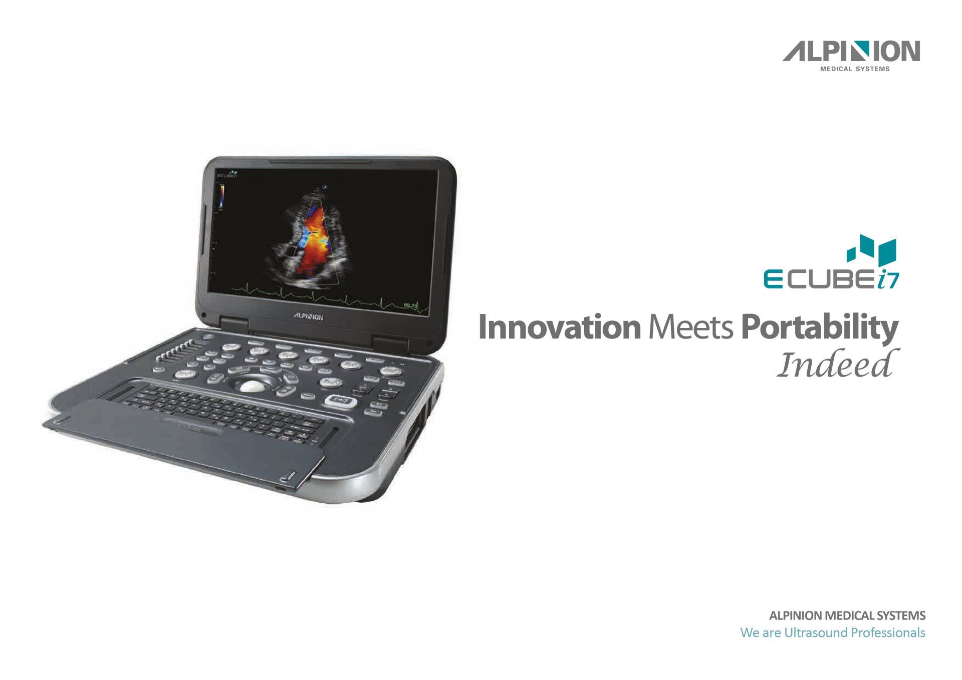
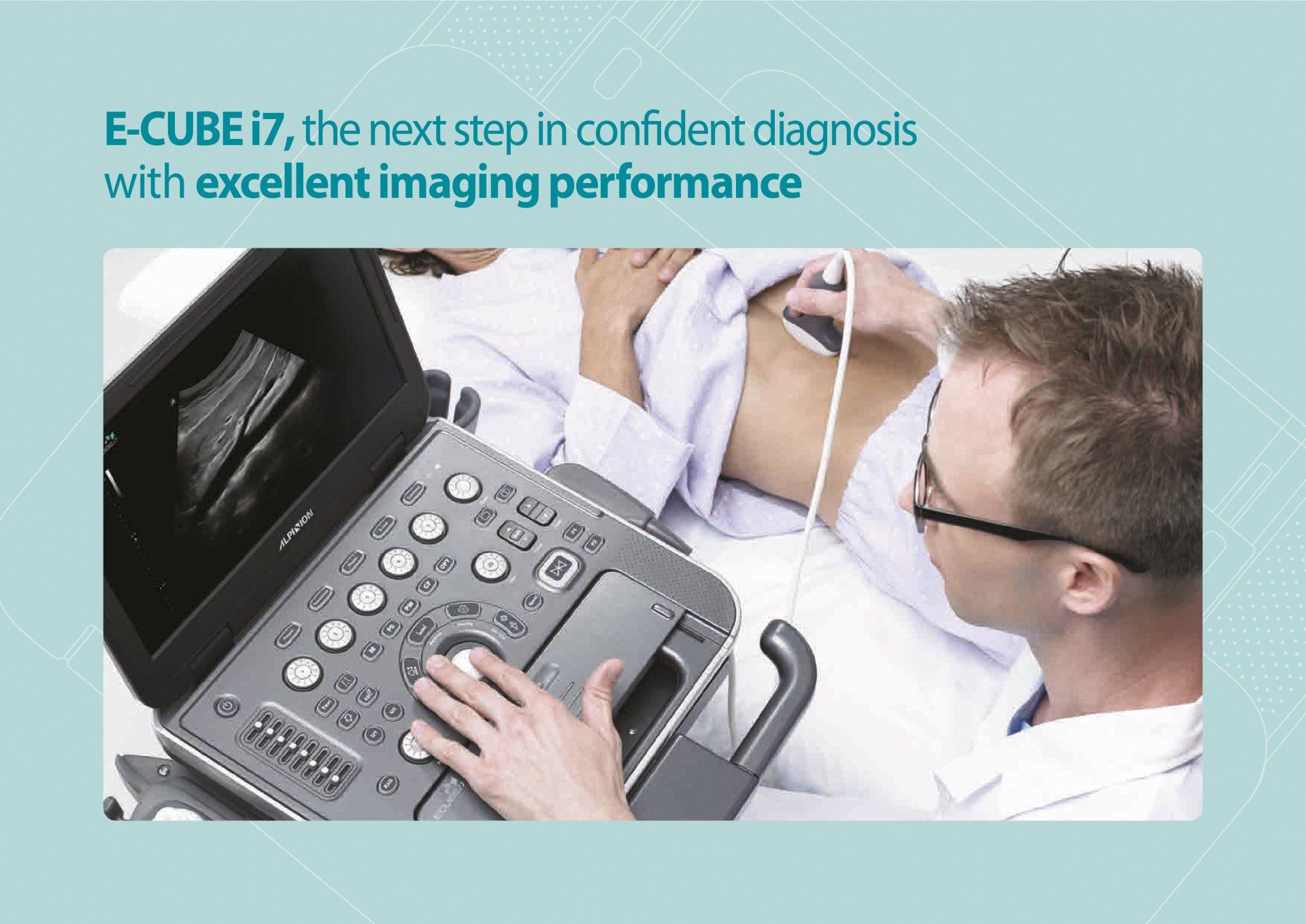
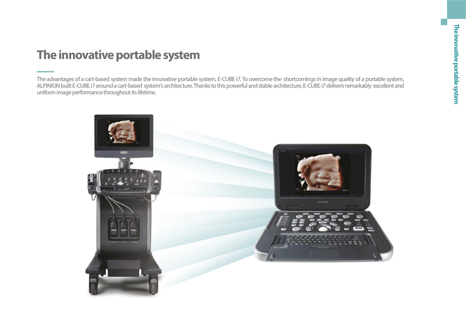
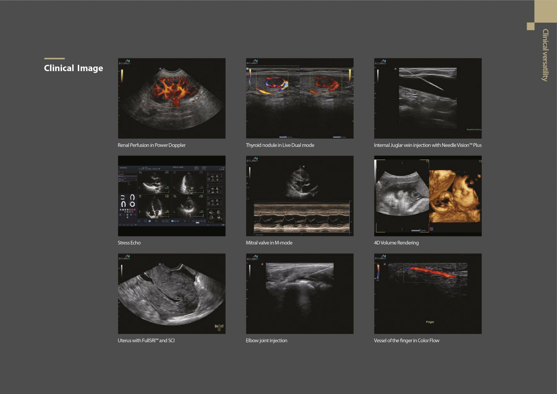
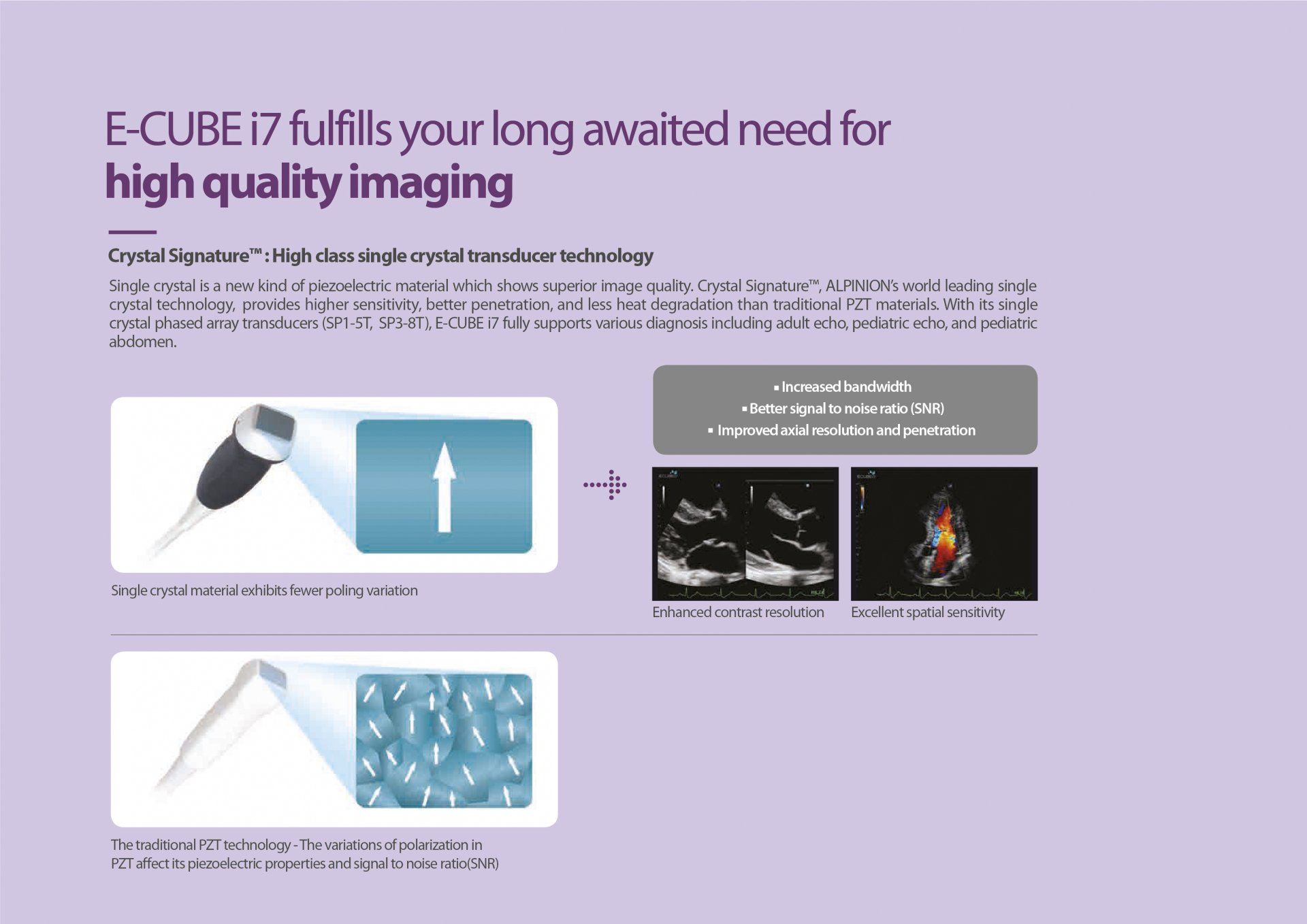
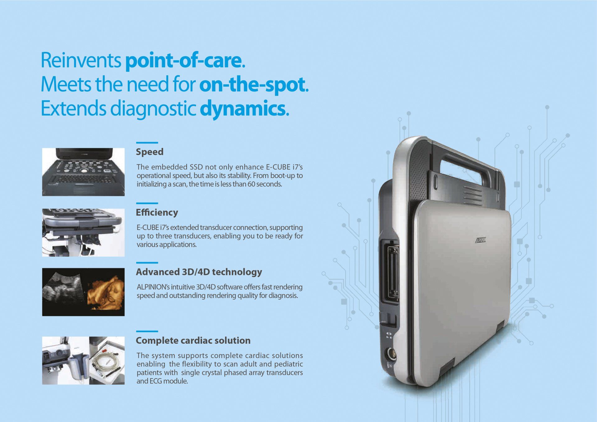
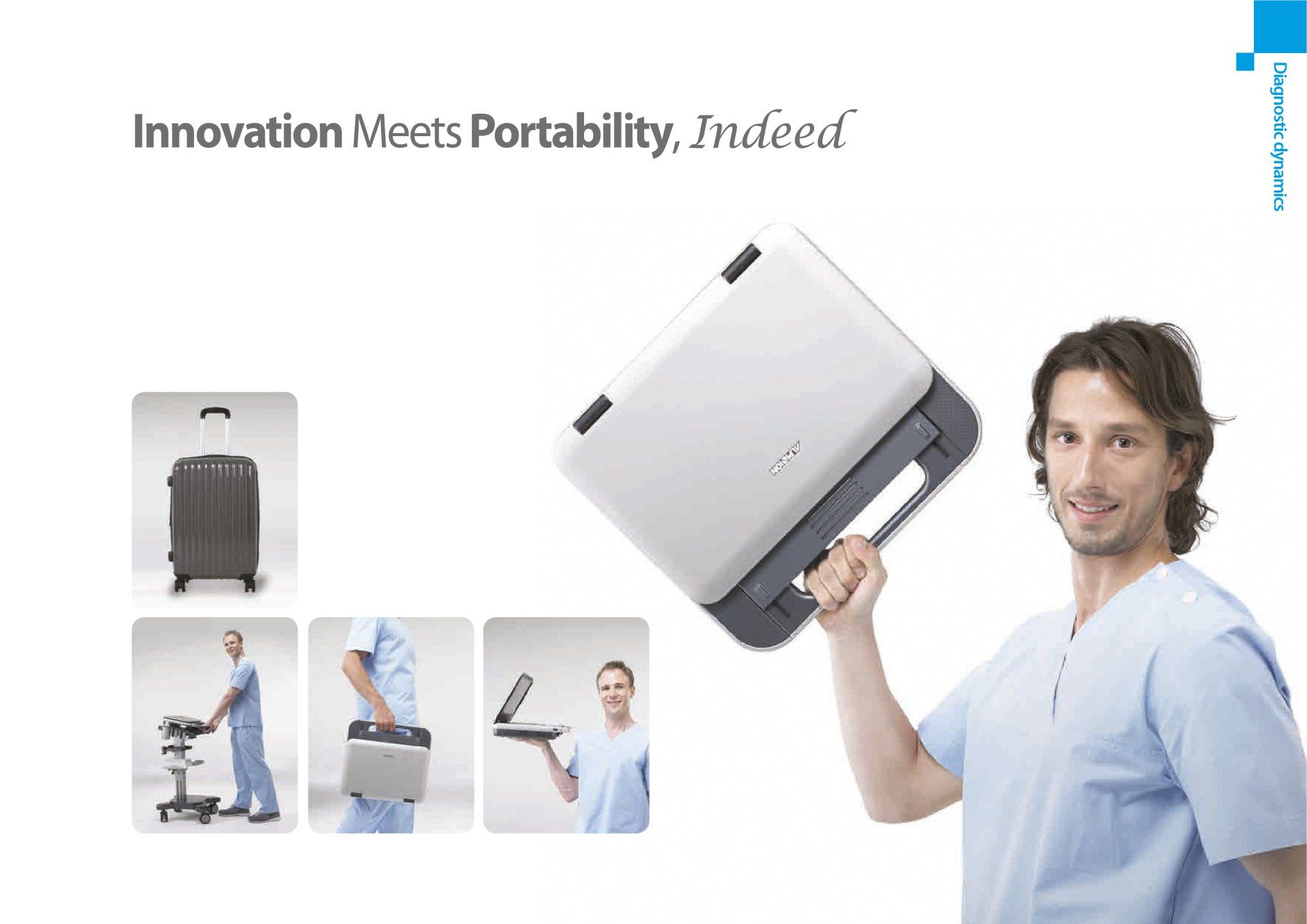
Echosonomovil uses the Alpinion E-Cube i7 equipment, a state-of-the-art compact system that allows obtaining optimal images in order to perform more accurate and reliable diagnoses, including complex cardiac studies.
INDICATIONS:
Ultrasound studies allow the detection of various diseases and also serve to guide invasive interventional procedures, such as aspiration biopsies, intra-articular injections, among others. It is also used to diagnose medical conditions associated with:
- pain
- inflammation and/or infection
- masses or tumors
- pregnancy
- various diseases
- pre and postoperative